
The reason tracing SACs is so exciting is that they connect to J cells. We want to start mapping this type of cell so we can see if/where the branches make contact with our two existing J cells.
Speaking of which, it's worth examining the two J cells we've done so far.
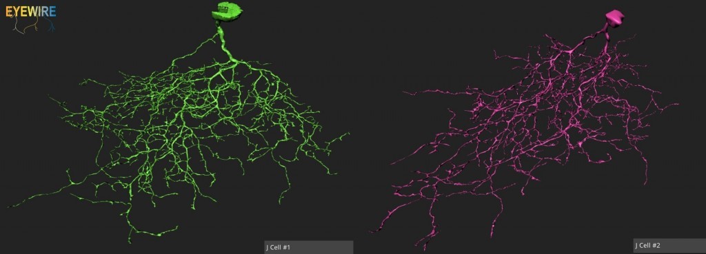
There's a definite likeness between the two, but it'd be interesting to look for differences between the two. You can look for differences here in the side by side photos, or you can go to the EyeWire Overview and switch between the two by clicking on the blue "change the cell" button. How similar do these cells look to you? What are the differences? Do you agree with us that these should be classified as the same type of cell?
Our blog post has more information and more images of the cells. So what do you guys think? Are these the same type of cell? Different? Let us know in the comments and please post screen shots so we can start the discussion.