
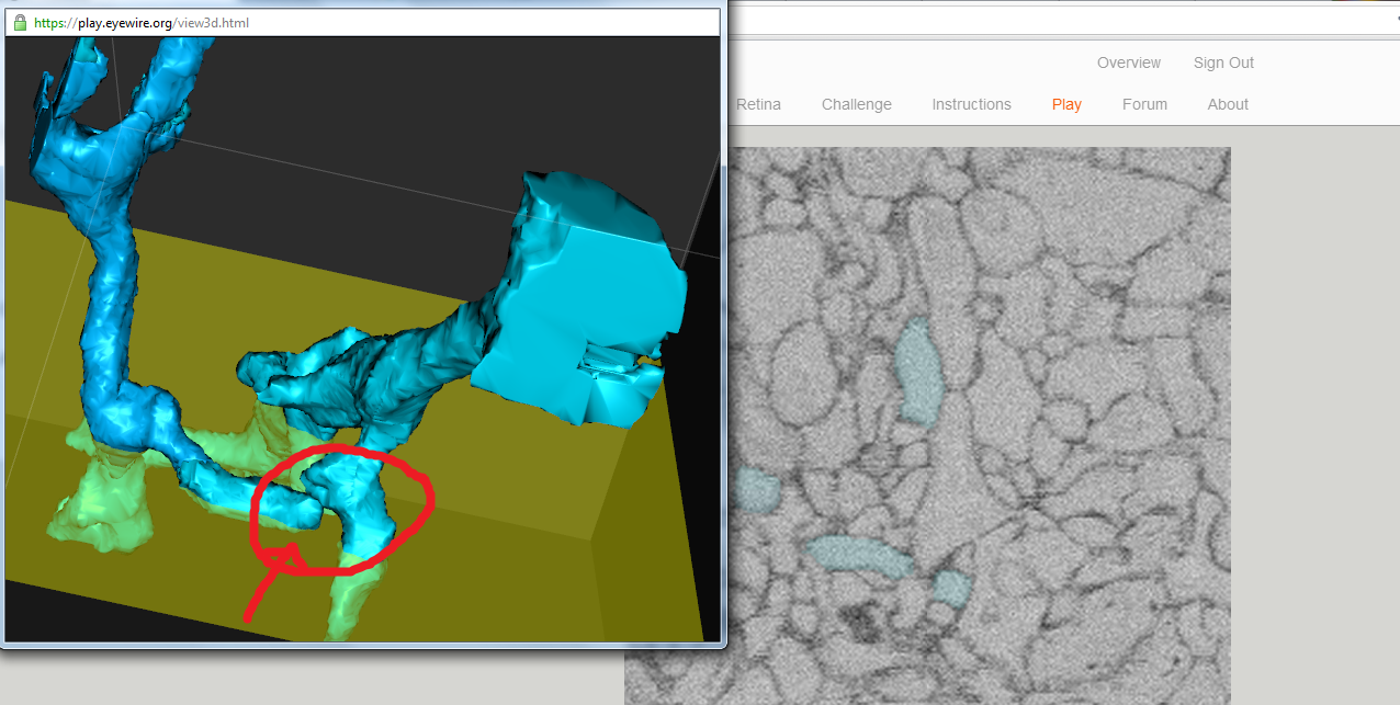
Sometimes you encounter two different branches which are very close together. My guess is these are the synapses, and you shouldn’t ‘fill them up’ as one? That’s at least the impression I got from the tutorial. What are the implications if you would submit them as depicted in the image?
This may or may not be a synapse, but I’d bet it’s not. This seems to me just an accidental contact of two neurons. Synapses have larger surface area of contact than this does, and the presynaptic sides have dented ‘holder’ on which postsynaptic sides can be placed.
We are reconstructing a neuron at a time. and you’re right, you should not fill these up as one. We didn’t design the system to distinguish different neurons from a single submission (nor no plan in that direction, at least for now). This kind of strategy is called as “sparse reconstruction” whereas what you would like to do is “dense reconstruction”. You could imagine pros and cons of them. Now we want to quickly get answers for what we are interested.
Another example. Visually it seems like they do connect at 1 or 2 slices, but my guess is that’s just due to the resolution not being high enough? Generally when adding a particular part results in an entire different ‘passing’ branch being added I don’t add it. I’m guessing this is what we should be doing, but it would help greatly to point this out in detail in the tutorial. Thank you!
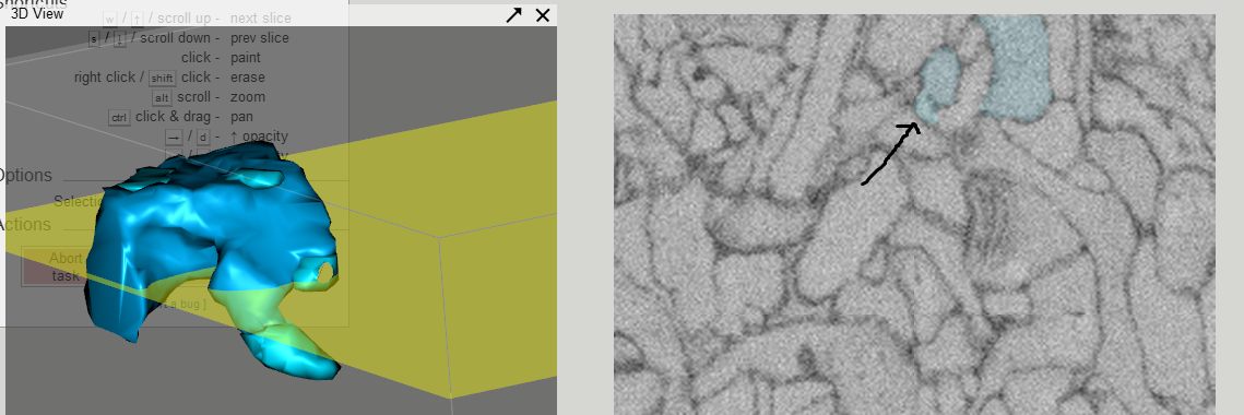

Another slice that makes me believe it is actually branching from the main structure. So … what are the guidelines on deciding on this?
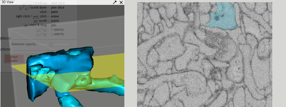
I agree with you. This seems like a branch from another neuron that is passing underneath of a π shaped structure. You had a good point. The overall shape of the structure that selected pieces make gives great hint.
The difficulty here is “staining gap”. A part (large or small) of cell membrane is not stained, and makes the boundary between two neurons disappear on a few slices. Decisions should be made based on larger context.
We are planning to prepare more training materials, and will certainly include these factors.
I am sometimes fooled by staining gaps also. Sometimes I scroll up and down a few times to see if the border only disappears for one or two slices. Also, one of the intuitions I have is that a neuron doesn’t form (or rarely forms) an X structure, which is what would happen if you had two separate sections merge into one section for a slice or two, and then split again. That’s helped me on more than one occasion to disambiguate an unclear slice.
Another intuition is that neurons rarely seem to get a very narrow waist in the middle of a branch. They might bulge momentarily, or they might get thin for a short run, but I’ve hardly seen a neuron get small momentarily.
Another intuition is that neurons don’t seem to form branches where the branches run together. They always seem to split off in different directions.
It’s difficult for me to tell without being able to scroll up and down through the volume. I do this rapidly back and forth to try to get a feel for where each path is going (or, as I like to say, where each path is blobbing off to).
I would say that as long as you see no cell membrane cross your blob, and that includes even a line which is mostly a gap, but has some “anchors” on the ends, as in your first image, then I’d say add it. If you end up violating some of the intuitions (neurons don’t form X’s, neurons don’t form parallel paths, neurons don’t end up with wasp-waists) then reconsider.
Thanks for posting screenshots, it makes it much easier for me to respond 

The example you posted is an example of a part of one cell wrapping around a branch from another cell. It’s really common. You’re also right to be suspicious when you add something that results in a new branch ‘passing’ through the original, don’t add the new branch. Thanks!