Is this a synapse?

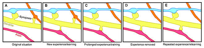
I believe I just found a synapse.


Although I can clearly make out a dendritic spine splitting off from the dendrite passing on the right, it’s not that clear whether it is connected to an axon.

Synapses always seem to be depicted with a dendritic spine touching a bigger section of the axon, as e.g. visible a bit higher up the axon.


Also, I only got 20 points for finishing this task. (I didn’t submit it with the dendrite attached.) Perhaps others often mistook it as being part of the axon?

It might be important giving a bit more background information in some extended tutorials to help in such scenarios.
I'm not sure I have already told you about this. There is a short description on how to discern synapses in this wiki page: http://wiki.eyewire.org/wiki/E2198
And Yeah, I think it is highly likely. 

Just one small correction: if the object on the left side is from the cell we are tracing, then it is the dendrite from ganglion cell, not an axon. The axons of ganglion cells are on other side of our dataset and we will scarcely have a chance to see them. then, the one on the right side should be the presynaptic partner of it. The synaptic boutons (presynpatic terminal) are depicted similarly as dendritic spines above. 


And regarding the low points, you don't get many scores for tasks that computer didn't make too much mistake. The biggest piece in the image is taking up most part of the branch and I'd assume that what you have added must be 2~3 small pieces at the bottom side of the image (around the location of your synapse). 

Thanks for the extra information! It’s great to be able to learn about this while actually seeing it when tracing.


I recall I did add quite a lot, which was why I was amazed by only 20 points. Usually the system gives 20 points when you did spend a considerable amount of time on the task, but nothing was added. Otherwise it gives e.g. 23 when little was added.

On an entirely different note. The ‘time’ measure is a bit vague, perhaps it’s better to replace it with ‘the amount of slices requested’.

E.g. If you have to scroll up and down three times, that indicates more ‘effort’ than only having to do one pass.


@whathecode The illustration that you posted is probably intended for the cerebral cortex.  Synapses between cortical pyramidal neurons are usually made onto dendritic spines, which is a classic fact found in most textbooks.  I’m not sure how many retinal synapses are made onto spines…we’d have to ask an expert or read some papers to find out.  I found this old paper by searching on Google Scholar, and I’m sure there are more. http://www.ncbi.nlm.nih.gov/pubmed/12106402     In any case, you’re right, this is a candidate for a synapse.