
Right now I’ve done less than 100 cubes but every once in a while I run across really odd shapes that make me question my judgement. Hopefully I am one of a minority of EyeWire starters that experience this. Maybe this can be the place in the forum where beginners can post their headscratcher pictures and receive comments from experienced users.
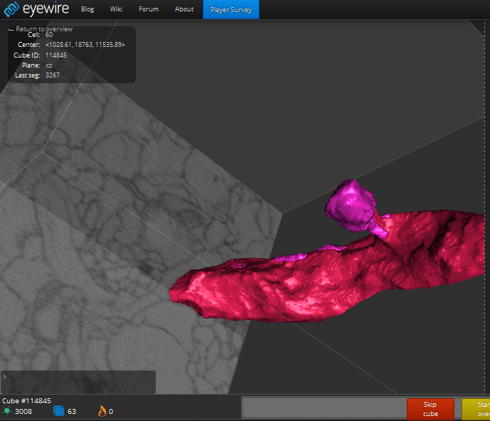
helge-#01:
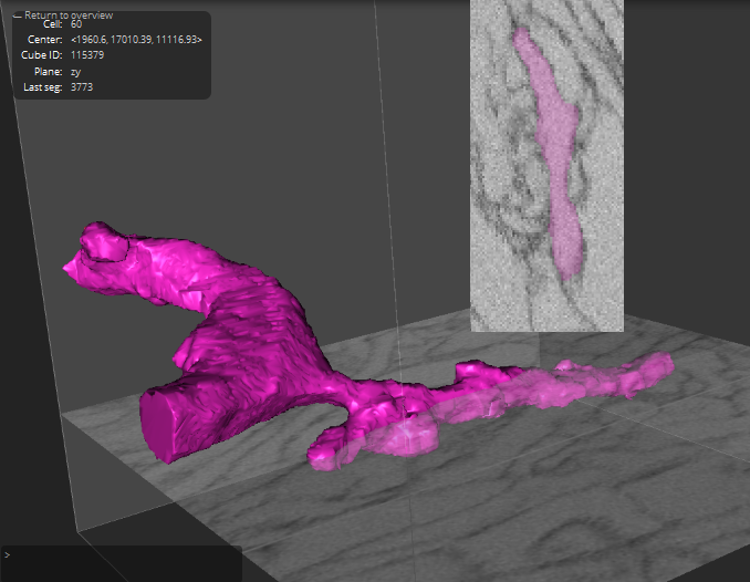
helge-#02:
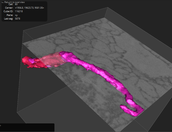
helge-#03:
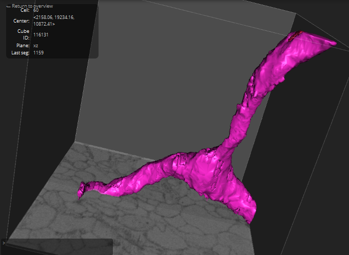
helge-#04:
please move/delete this thread if you find it inappropriate or there is already a different place for submissions.
ps. how about a “submit a snapshot” function for views/cubes to be reviewed, combined with user galleries that contain the image plus classification/comment after review?
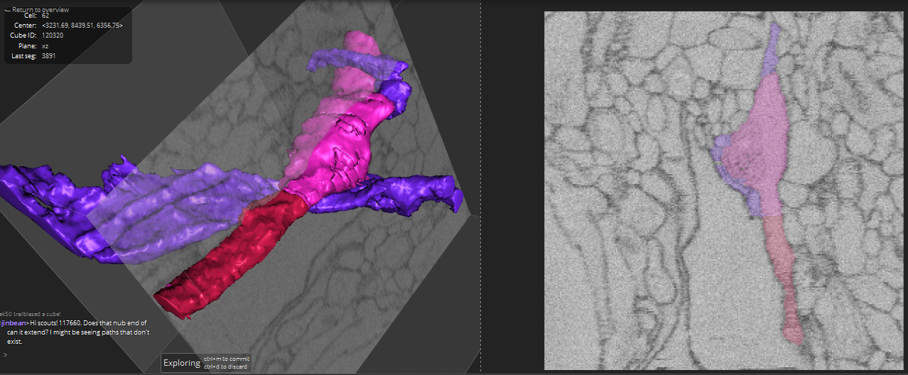

 If you didn’t add the blue explore-mode part to cube
 C
C
