Hi there!
Do you have a science question about EyeWire or the brain in general? Ask here and you’ll get an answer from HQ. The best questions may even end up on the EW blog 
Amy

Hi there!
Do you have a science question about EyeWire or the brain in general? Ask here and you’ll get an answer from HQ. The best questions may even end up on the EW blog
Amy
This topic is now a banner. It will appear at the top of every page until it is dismissed by the user.
This topic is no longer a banner. It will no longer appear at the top of every page.
Q&A from 신경 과학 관련 질문 (한글 포스팅, Questions in Korean about neuroscience)
@scoobi’s question: 신경 과학 관련 질문 (한글 포스팅, Questions in Korean about neuroscience)
Cells in EyeWire are all from mouse’ retina. How similar a person’s retina and a mouse’s are from each other? Isn’t there any neuron that exclusively exists in either being’s retina? In addition, I am curious to know that there are such special neurons which only exist in a specific
species.
@jinseop’s answer: 신경 과학 관련 질문 (한글 포스팅, Questions in Korean about neuroscience)
It is a good question. Like other organs in body, retina is more similar among genetically close species and more different among genetically farther species. Mice and human beings are relatively close species and structures of their retinas are similar.
It is common to animals like mammals and reptiles that their retina has five different categories of neurons – photoreceptor cells, horizontal cells, bipolar cells, amacrine cells, and ganglion cells – and they form layered structure. It is believed that the photoreceptors and horizontal cells have little differences among species. Although the bipolar cells, amacrine cells, and ganglion cells have not been completely analyzed in any of species yet, 8 - 15 types of photoreceptor cells, 50 -100 types of amacrine cells, and 30 - 50 type of ganglion cells are expected to be found from different species. We assume 50 - 80% of those cells are shared between mouse and human retina. The word “shared” here means that we can find anatomically (morphologically) identical counterpart of a cell from the other retina. However, more study is needed to verify that the shared cells are genetically and functionally identical in both retina.
While the basic visual functionality of retina should be common to any animal, the retina of different species must differentiate to meet the biological necessities specific to the species. For example, preys must be good at recognizing their predators; predators must be good at recognizing their preys. Those differentiated retinas end up having different set of cell types and different connectome.
Do we know what type/kind of cell is CAND cand 1439 jump ID: 947221 and are we going to trace it? It looks AWESOME! 
Are the cells we complete in eyewire checked or edited after in OMNI? For fused mergers we could not remove in ew or other such problems?
Did we ever find out what kind of cell it was?
During the countdown to neuropia and now afterwards in the bonus cells, we’ve found and traced aprox 8-10 starburst cells. I imagine all those are from the ganglion layer? Is it strange/new to find SBs in the ganglion layer or is it already a phenomenon that has been previously recorded and explained?
Hi!
You give us eyewirer’s many scientific information about what we (and you) do.
That’s great!
I’d like to understand a bit more detailed what kind of cells we are tracing,
thus I’d like to be able to roughly identify cell types. I looked at the
EW-museum and the many blogs and searched in internet about retinal cell types
etc.
So I have some questions:
Dataset ranges from Ganglion-cell-layer to about middle of INL (see attachment
vertebrate retina)? So if we get a cell with cellbody, we can say, it is either
a ganglion-cell or an amacrine-cell, and it cannot be a Bipolar (their
cellbodies being outside dataset)?
What’s the difference in Dataset range between Neuropia and Dig? Dig going a
bit farther into INL?
If amacrine, cellbody can be located in GC-layer or in INL? If yes, we
eyewirers cannot see in which layer the cellbody is located, can we? (When we
choose a cell, how is it oriented as compared to the dataset?) Were Starbursts
coming from GC-Layer or INL?
What kind of amacrine cell is All? Shape outlined in draft of “vertebrate
retina” looks quite special, did we ever trace such a cell? Reminds me a bit of
shape of cells like Relic 1.100.
Why in museum no amacrine cells are represented (or are some under GC’s)?
Bipolars are well represented in the museum, and numbering quite
comprehensible. I miss though the link to cell-numbers we eyewirers knew. And
the link to cell-names as RBC and CBC (are there more?)
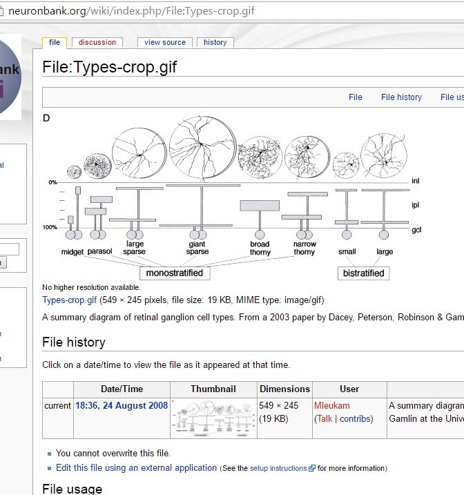
Numbering of Ganglion cells in the museum is harder to understand with all
these abbreviations
Here too I miss the links. There seem to be many different types of
ganglion-cells. I tried to relate them with cell-types from publications (see
attachment GC-Layer cell types and link http://www.jneurosci.org/content/22/9/
3831.full.pdf).
(gave up, and obviously characterization is going on).
P-type, Midget, Beta = GC’s we mostly traced? (Aside bistratified)
M-type, parasol. Alpha = ? Have an example in the museum? (sounds like
amacrine)
large-sparse (in other publication G8) = Artifact 1.88?
thorny GCs = cells looking like Relic 1.100 ?
How you name these cell-types?
Couldn’t you give some more information in the museum-site and/or a summing-up
of cell-groups in there? (Perhaps you do not want to give too many infos for
public as long data are not published, I can understand)
Is there a link-list of cell-numbers, cell-names we players know and the number
in museum?
You see, I’m quite overstrained. Perhaps you can give me some help. I’m aware,
it won’t help much science, but eyewire is more than a game for me, and I’d
like to understand as much as possible.
If the goal is tracing cell interactions why not just draw the best line along axons? Why fill it out immediately? For instance, I’ve seen an ImageJ module that automates filling in the axon from a line.
Hi Mike,
It sounds like you’re talking about a type of reconstruction method called skeleton tracing (also known as skeletonization or topological skeleton). Skeleton tracing is where you place a dot every few slides in the middle of the branch you are tracing - the computer fills in the rest by extruding a shape along the line of dots.
Eyewire and the Seung lab use full reconstruction. We’ve found that full reconstruction tends to be more accurate than skeleton tracing (small branches can be more easily missed in skeleton tracing). It also provides us with a full surface texture and structure of the neuron. We’re looking for synapses and examining the synaptic connection between cells. Synapses can be better found when we can see the full structure as it looks like a “handshake” between two cells - looking for nubs helps us identify actual synapses vs an “incidental” contact between cells.
Hope that helps explain things!
I read awhile back that the research shows signs that might help understand/prevent Alzheimer. Has there been any updates on this? I understand “shows signs” could mean 80 years down the road or it could be 80 days. Forgive me, I lack the ability to fulling word what I’m trying to say, but thank you for reading and answering if you do.
Hi deadxdying,
Eyewire’s current dataset deals with the retina of the eye and understanding the neural circuits related to primarily vision so our research doesn’t currently deal with Alzheimer’s disease. Eyewire is one the first steps being taken towards improving brain mapping and creating connectomes. The field of connectomics certainly has far-reaching applications including better understanding Alzheimer’s.
Perhaps you have read a recent article talking about our involvement with the WeCureALZ project. Eyewire is beginning preliminary work with WeCureALZ to adapt their dataset for a new game platform. The above article also has some interesting updates on recent findings for Alzheimer’s and I suggest you check out the WeCureALZ project.